Images & Videos: Hip Ultrasound
Items 1 to 5 of 5 - Click images to view larger
Ultrasound equipment
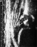
Ultrasound image of a hip in a six month old baby. The arrows point to the early bone formation within the head of the femur.
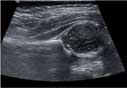
Ultrasound image of an infant hip. Arrows point to a round cartilage structure representing the head of the femur sitting in the joint space (ball in socket).
5 month old female: hip evaluated for congenital dysplasia
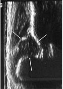
Ultrasound showing a dislocated hip--head of femur out of alignment with hip socket.
Ultrasound showing a dislocated hip--head of femur out of alignment with hip socket.
5 month old female: hip evaluated for congenital dysplasia
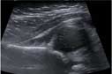
Ultrasound of a normal hip.
a. gluteus muscle
b. ilium
c. acetabulum
d. head of femur
Ultrasound of a normal hip.
a. gluteus muscle
b. ilium
c. acetabulum
d. head of femur