Images & Videos: Chest X-ray
Items 1 to 10 of 33 - Click images to view larger
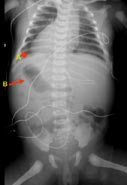
In this x-ray image, the letter A points to the dextrocardia (displacement of the heart to the right). The letter B points to the stomach in the right upper abdominal quadrant. This is situs inversus (reversal of position) totalis.
In this x-ray image, the letter A points to the multiple gas bubbles in the left hemithorax. This is a congenital diaphragmatic hernia.
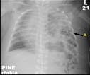
Frontal radiograph of the chest demonstrating a right tension pneumothorax.
Radiography equipment
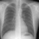
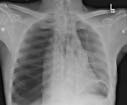
Chest x-ray. Frontal view of a male patient.
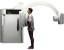
 A radiologic technologist preparing a patient for a chest x-ray. A chest x-ray produces images of the heart, lungs, airways, blood vessels and the bones of the spine and chest.
 Your Radiologist Explains Chest X-ray
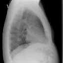
Chest x-ray. Side view or lateral view of the chest.