Images & Videos: Renal Scintigraphy
Items 1 to 10 of 10 - Click images to view larger
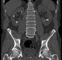
Computed tomography (CT) scan of a patient’s kidneys showing kidney stones (arrows). For more information see Kidney and Bladder Stones.
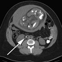
Axial computed tomography (CT) image from a renal (kidney) exam in a pregnant patient showing a stone in the middle segment of the right ureter (arrow). For more information see Kidney and Bladder Stones.
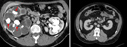
Left = Computed tomography (CT) image showing large stones (arrows) and kidney scarring. Right = Computed tomography (CT) image showing normal kidneys.
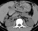
Multi-detector computed tomography (MDCT) scan of a pediatric patient showing a right renal (kidney) stone. For more information see Kidney and Bladder Stones.
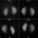
Four images from a normal cortical renal scintigraphy.
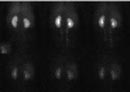
Dynamic images from a normal functional renal imaging.
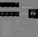
Normal graphs and split function from functional renal imaging.
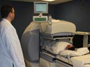
Photograph of a technologist performing a renal scan on a patient using a gamma camera.
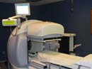
Photograph of a dual headed SPECT-CT gamma camera.
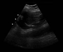
Ultrasound image showing a kidney stone (arrow) obstructing urine flow. For more information see Ultrasound and Kidney and Bladder Stones.