Images & Videos: Interventional Radiology (IR)
Items 1 to 10 of 113 - Click images to view larger
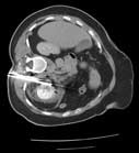
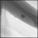
Radiofrequency ablation of a renal tumor: Cluster radiofrequency probe is positioned in the patient's renal cell carcinoma. For more information see Radiofrequency Ablation of Kidney Tumors.
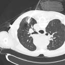
During radiofrequency ablation, electrodes are placed in the patient's lung cancer using CT guidance. For more information see Radiofrequency Ablation of Lung Tumors.
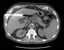
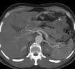
CT-guided radiofrequency ablation of a liver tumor: A cluster electrode is positioned in the patient's hepatocellular carcinoma. For more information see Radiofrequency Ablation of Liver Tumors.
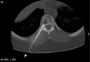
CT-guided needle biopsy of a vertebral body lesion.
CT image is shown with needle (bright white line) placed in vertebral body and needle tip in a lesion in the right-sided portion of the vertebral body.
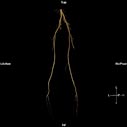
Normal three-dimensional CT angiogram of the lower extremities. For more information see CT Angiography (CTA).
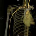
Normal three-dimensional (3-D) CT angiogram of the right upper arm shows major arm artery and relationship to neighboring bones/rib cage. For more information see CT Angiography (CTA).

Reformatted image of the mid-abdomen creating a 'slab' showing the liver and intestines. The abdominal aorta and branches going to the liver, spleen and stomach are visible. This view is looking up toward the head of the patient.
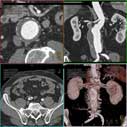
3-D images of the abdominal aorta showing the main vessels in the abdomen.