Images & Videos: Appendicitis
Items 1 to 7 of 7 - Click images to view larger
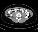
Appendicitis: A CT cross-section through the patient's lower abdomen has also cut through the swollen, inflamed appendix in two places (arrows). Inflammation has spread to the nearby intra-abdominal fat (arrowhead). For more information see Appendicitis.
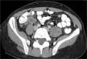
Appendicitis: The appendix (A) is distended and inflamed. In this patient the appendix has not yet ruptured. For more information see Appendicitis.
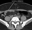
CT scan of a normal appendix in the right lower abdomen. The appendix normally connects with the right colon and contains air (this appears black on the scan). Air in the appendix excludes appendicitis since this means that the appendix is not obstructed or inflamed. For more information see Appendicitis.
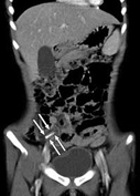
Coronal computed tomography (CT) scan of a pediatric patient with acute appendicitis and appendicolith (inflammation of the appendix) showing a calcified (hardened) lesion (arrows). For more information see Appendicitis.
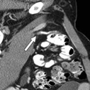
Intravenous (IV) contrast-enhanced computed tomography (CT) image in patient with acute appendicitis shows contrast material opacification of the appendiceal lumen (arrow). For more information see Appendicitis.