Images & Videos: CAT Scan (CT) - Head
Items 11 to 20 of 29 - Click images to view larger
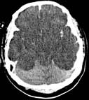
Axial non-contrast CT scan demonstrates relative hyperdensity of the cerebellum in relation to the supratentorial brain due to diffuse anoxic (absence of oxygen) injury.
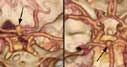
Two different views of CT brain angiogram reveal an aneurysm (arrows) arising from the basilar artery.

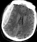
Recent bleeding (subdural hematoma) in an injured patient is seen as a bright mass that is pushing the brain to the other side.

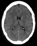
Perfusion CT in a patient with stroke demonstrates the part of the brain with severely decreased blood flow (arrows).
A CT scan of the head.

Dr. Robert Zimmerman discusses head trauma in children.
A CT scan of the head showing a large blood clot after a head injury - epidural hematoma.
 Your Radiologist Explains The use of Contrast Material for Head CT Scanning
 Your Radiologist Explains CT of the Head

Mandibular (jaw) fracture. A 3-D surface rendered image from CT clearly depicts left-sided condyloid process fracture. The 3-D rendering is often requested by plastic surgery for treatment planning.