Images & Videos: Chest X-ray
Items 11 to 20 of 31 - Click images to view larger
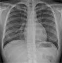
Posteroanterior chest on a normal female.
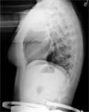
Lateral chest on a normal female.
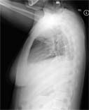
Lateral chest on someone with pneumonia. The pneumonia is the white substance in the lower part of each lung.
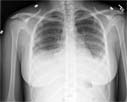
Posteroanterior chest on someone with pneumonia. The pneumonia is the white substance in the lower part of each lung.
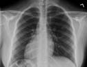
An x-ray image showing perihilar lymphadenopathy in a patient with sarcoidosis.
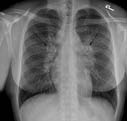
A single posterior-anterior radiographic image (x-ray) of the chest demonstrating the heart situated in the right chest, otherwise known as dextrocardia.
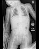
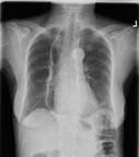
Kiddiegram of a child showing chest and abdomen.
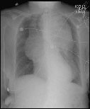
Chest x-ray demonstrates a large thoracic aortic aneurysm as the source of a female patient's pain.
A frontal radiograph of the chest demonstrating a very dilated, air filled esophagus in a patient with achalasia.
 A radiologic technologist preparing a pediatric patient for a chest x-ray. A chest x-ray produces images of the heart, lungs, airways, blood vessels and the bones of the spine and chest.