Images & Videos: Bone X-ray
Items 21 to 30 of 43 - Click images to view larger
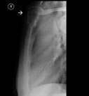
X-ray of the upper frontal chest wall of a patient with cancer. The sternum (breastbone) has an expanded appearance which is due to a destructive lesion.

X-ray showing an osteosarcoma of the femur.

X-ray of the long bone in the thigh demonstrates calcified medullary bone infarcts. Patients with medullary infarcts are usually asymptomatic.

X-ray of a child with rickets demonstrating typical bowing of the legs.
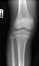
A normal knee in a 12 year old (anterior-posterior view).
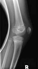
A normal knee in a 12 year old (lateral view).
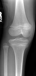
A normal knee in a 12 year old (oblique view).

Normal appearance of the lumbosacral spine
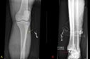
Frontal (A) and lateral (B) radiographs of the left leg and ankle demonstrate a fracture of the distal tibia (yellow arrow) along with a fracture of the proximal fibula (red arrow) with separation of the long bones (tibia and fibula) consistent with a Maissoneuve fracture.
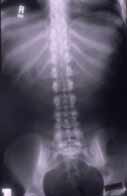
X-ray showing lumbar spine (front view)