Ultrasound - Musculoskeletal

- What is Ultrasound Imaging of the Musculoskeletal System?
- What are some common uses of the procedure?
- How should I prepare?
- What does the equipment look like?
- How does the procedure work?
- How is the procedure performed?
- What will I experience during and after the procedure?
- Who interprets the results and how do I get them?
- What are the benefits vs. risks?
- What are the limitations of Ultrasound Imaging of the Musculoskeletal System?
What is Ultrasound Imaging of the Musculoskeletal System?

Ultrasound is safe and painless, and produces pictures of the inside of the body using sound waves. Ultrasound imaging, also called ultrasound scanning or sonography, involves the use of a small transducer (probe) and ultrasound gel placed directly on the skin. High-frequency sound waves are transmitted from the probe through the gel into the body. The transducer collects the sounds that bounce back and a computer then uses those sound waves to create an image. Ultrasound examinations do not use ionizing radiation (as used in x-rays), thus there is no radiation exposure to the patient. Because ultrasound images are captured in real-time, they can show the structure and movement of the body's internal organs, as well as blood flowing through blood vessels.
Ultrasound imaging is a noninvasive medical test that helps physicians diagnose and treat medical conditions.
Ultrasound images of the musculoskeletal system provide pictures of muscles, tendons, ligaments, joints and soft tissue throughout the body.
What are some common uses of the procedure?

Ultrasound images are typically used to help diagnose:
- tendon tears, or tendinitis of the rotator cuff in the shoulder, Achilles tendon in the ankle and other tendons throughout the body.
- muscle tears, masses or fluid collections.
- ligament sprains or tears.
- inflammation or fluid (effusions) within the bursae and joints.
- early changes of rheumatoid arthritis.
- nerve entrapments such as carpal tunnel syndrome.
- benign and malignant soft tissue tumors.
- ganglion cysts.
- hernias.
- foreign bodies in the soft tissues (such as splinters or glass).

- dislocations of the hip in infants.
- fluid in a painful hip joint in children.
- neck muscle abnormalities in infants with torticollis (neck twisting).
- soft tissue masses (lumps/bumps) in children.
How should I prepare?
You should wear comfortable, loose-fitting clothing for your ultrasound exam. You may need to remove all clothing and jewelry in the area to be examined.
You may be asked to wear a gown during the procedure.
Ultrasound examinations are very sensitive to motion, and an active or crying child can prolong the examination process. To ensure a smooth experience, it often helps to explain the procedure to the child prior to the exam. Bringing books, small toys, music or games can help to distract the child and make the time pass quickly. The ultrasound exam room may have a television. Feel free to ask for your child's favorite channel.
No other preparation is required.
What does the equipment look like?
Ultrasound scanners consist of a console containing a computer and electronics, a video display screen and a transducer that is used to do the scanning. The transducer is a small hand-held device that resembles a microphone, attached to the scanner by a cord. The transducer sends out inaudible high frequency sound waves into the body and then listens for the returning echoes from the tissues in the body. The principles are similar to sonar used by boats and submarines.
The ultrasound image is immediately visible on a video display screen that looks like a computer or television monitor. The image is created based on the amplitude (loudness), frequency (pitch) and time it takes for the ultrasound signal to return from the area of the patient being examined to the transducer (the device used to examine the patient), as well as the type of body structure and composition of body tissue through which the sound travels. A small amount of gel is put on the skin to allow the sound waves to travel back and forth from the transducer.
How does the procedure work?
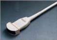
Ultrasound transducer
Ultrasound imaging is based on the same principles involved in the sonar used by bats, ships and fishermen. When a sound wave strikes an object, it bounces back, or echoes. By measuring these echo waves, it is possible to determine how far away the object is as well as the object's size, shape and consistency (whether the object is solid or filled with fluid).
In medicine, ultrasound is used to detect changes in appearance, size or contour of organs, tissues, and vessels or detect abnormal masses, such as tumors.
In an ultrasound examination, a transducer both sends the sound waves and receives the echoing waves. When the transducer is pressed against the skin, it directs small pulses of inaudible, high-frequency sound waves into the body. As the sound waves bounce off internal organs, fluids and tissues, the sensitive microphone in the transducer records tiny changes in the sound's pitch and direction. These signature waves are instantly measured and displayed by a computer, which in turn creates a real-time picture on the monitor. One or more frames of the moving pictures are typically captured as still images. Small loops of the moving “real time” images may also be saved.
How is the procedure performed?

For certain ultrasound examinations of the musculoskeletal system, the patient may be seated on an examination table or a swivel chair. For other ultrasound exams, the patient is positioned lying face-up or face-down on an examination table. The radiologist or sonographer may ask you to move the extremity being examined or may move it for you to evaluate the anatomy and function of the joint, muscle, ligament or tendon.
Most ultrasound studies of infants and children are performed with the child lying on his or her back on the examination table, but other positions may be required.
After you are positioned on the examination table, the radiologist or sonographer will apply a warm water-based gel to the area of the body being studied. The gel will help the transducer make secure contact with the body and eliminate air pockets between the transducer and the skin that can block the sound waves from passing into your body. The transducer is placed on the body and moved back and forth over the area of interest until the desired images are captured.
There is usually no discomfort from pressure as the transducer is pressed against the area being examined. However, if scanning is performed over an area of tenderness, you may feel pressure or minor pain from the transducer.
Once the imaging is complete, the clear ultrasound gel will be wiped off your skin. Any portions that are not wiped off will dry to a powder. The ultrasound gel does not stain or discolor clothing.
What will I experience during and after the procedure?
Ultrasound examinations are painless and easily tolerated by most patients.
Musculoskeletal ultrasound examination is usually completed within 15 to 30 minutes but may occasionally take longer.
When the examination is complete, you may be asked to dress and wait while the ultrasound images are reviewed.
After an ultrasound examination, you should be able to resume your normal activities immediately.
Who interprets the results and how do I get them?
A radiologist, a physician specifically trained to supervise and interpret radiology examinations, will analyze the images and send a signed report to your primary care physician, or to the physician or other healthcare provider who requested the exam, and he/she will share the results with you. In some cases the radiologist may discuss results with you at the conclusion of your examination.
Follow-up examinations may be necessary, and your doctor will explain the exact reason why another exam is requested. Sometimes a follow-up exam is done because a suspicious or questionable finding needs clarification with additional views or a special imaging technique. A follow-up examination may also be necessary so that any change in a known abnormality can be monitored over time. Follow-up examinations are sometimes the best way to see if treatment is working or if an abnormality is stable over time.
What are the benefits vs. risks?
Benefits
- Most ultrasound scanning is noninvasive (no needles or injections).
- Occasionally, an ultrasound exam may be temporarily uncomfortable, but it is almost never painful.
- Ultrasound is widely available, easy-to-use and less expensive than other imaging methods.
- Ultrasound imaging is extremely safe and does not use any ionizing radiation.
- Ultrasound scanning gives a clear picture of soft tissues that do not show up well on x-ray images.
- Ultrasound provides real-time imaging, making it a good tool for guiding minimally invasive procedures such as needle biopsies and fluid aspiration.
- Patients with cardiac pacemakers and certain types of metallic implants or fragments in the body cannot be safely exposed to the strong magnetic field required for magnetic resonance imaging (MRI); however, patients can safely receive ultrasound imaging.
- Ultrasound is also an excellent alternative to MRI for claustrophobic patients.
- Compared to MRI, ultrasound may provide greater internal detail when assessing soft tissue structures such as tendons and nerves.
- Because ultrasound images are captured in real time, they can show the movement of a soft tissue structure such as a tendon, joint or an extremity.
- Ultrasound imaging is faster than MRI and does not require the patient to remain completely still, allowing infants to be imaged without sedation.
- The hip joints of infants, unlike those of adults, are largely made of cartilage. Ultrasound is able to clearly see cartilage.
Risks
- For standard diagnostic ultrasound, there are no known harmful effects on humans.
What are the limitations of Ultrasound Imaging of the Musculoskeletal System?
Ultrasound has difficulty penetrating bone and, therefore, can only see the outer surface of bony structures and not what lies within (except in infants who have more cartilage in their skeletons than older children or adults). For visualizing internal structure of bones or certain joints, other imaging modalities such as MRI are typically used.
There are also limitations to the depth that sound waves can penetrate; therefore, deeper structures in larger patients may not be seen easily.
Ultrasound has not proven useful in detecting whiplash injuries or most other causes of back pain.
Locate an ACR-accredited provider: To locate a medical imaging or radiation oncology provider in your community, you can search the ACR-accredited facilities database.
This website does not provide costs for exams. The costs for specific medical imaging tests and treatments vary widely across geographic regions. Many—but not all—imaging procedures are covered by insurance. Discuss the fees associated with your medical imaging procedure with your doctor and/or the medical facility staff to get a better understanding of the portions covered by insurance and the possible charges that you will incur.
Web page review process: This Web page is reviewed regularly by a physician with expertise in the medical area presented and is further reviewed by committees from the American College of Radiology (ACR) and the Radiological Society of North America (RSNA), comprising physicians with expertise in several radiologic areas.
Outside links: For the convenience of our users, RadiologyInfo.org provides links to relevant websites. RadiologyInfo.org, ACR and RSNA are not responsible for the content contained on the web pages found at these links.
Images: Images are shown for illustrative purposes. Do not attempt to draw conclusions or make diagnoses by comparing these images to other medical images, particularly your own. Only qualified physicians should interpret images; the radiologist is the physician expert trained in medical imaging.
This page was reviewed on July 16, 2013