Intravenous Pyelogram (IVP)
- What is an Intravenous Pyelogram (IVP)?
- What are some common uses of the procedure?
- How should I prepare?
- What does the equipment look like?
- How does the procedure work?
- How is the procedure performed?
- What will I experience during and after the procedure?
- Who interprets the results and how do I get them?
- What are the benefits vs. risks?
- What are the limitations of IVP exams?
What is an Intravenous Pyelogram (IVP)?
An intravenous pyelogram (IVP) is an x-ray examination of the kidneys, ureters and urinary bladder that uses iodinated contrast material injected into veins.
An x-ray (radiograph) is a noninvasive medical test that helps physicians diagnose and treat medical conditions. Imaging with x-rays involves exposing a part of the body to a small dose of ionizing radiation to produce pictures of the inside of the body. X-rays are the oldest and most frequently used form of medical imaging.
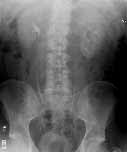
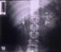
When a contrast material is injected into a vein in the patient's arm, it travels through the blood stream and collects in the kidneys and urinary tract, turning these areas bright white on the x-ray images. An IVP allows the radiologist to view and assess the anatomy and function of the kidneys, ureters and the bladder.
What are some common uses of the procedure?
An intravenous pyelogram examination helps the radiologist assess abnormalities in the urinary system, as well as how quickly and efficiently the patient's system is able to handle fluid waste.
The exam is used to help diagnose symptoms such as blood in the urine or pain in the side or lower back.
The IVP exam can enable the radiologist to detect problems within the urinary tract resulting from:
- kidney stones
- enlarged prostate
- tumors in the kidney, ureters or urinary bladder
- surgery on the urinary tract
- congenital anomalies of the urinary tract
How should I prepare?
Your doctor will give you detailed instructions on how to prepare for your IVP study.
You will likely be instructed not to eat or drink after midnight on the night before your exam. You may also be asked to take a mild laxative (in either pill or liquid form) the evening before the procedure.
You should inform your physician of any medications you are taking and if you have any allergies, especially to iodinated contrast materials. Also inform your doctor about recent illnesses or other medical conditions.
You may be asked to remove some or all of your clothes and to wear a gown during the exam. You may also be asked to remove jewelry, removable dental appliances, eye glasses and any metal objects or clothing that might interfere with the x-ray images.
Women should always inform their physician and x-ray technologist if there is any possibility that they are pregnant. Many imaging tests are not performed during pregnancy so as not to expose the fetus to radiation. If an x-ray is necessary, precautions will be taken to minimize radiation exposure to the baby. See the Safety page (www.RadiologyInfo.org/en/safety/) for more information about pregnancy and x-rays.
What does the equipment look like?
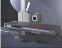
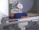
The equipment typically used for this examination consists of a radiographic table, one or two x-ray tubes and a television-like monitor that is located in the examining room. Fluoroscopy, which converts x-rays into video images, is used to watch and guide progress of the procedure. The video is produced by the x-ray machine and a detector that is suspended over a table on which the patient lies.
How does the procedure work?
X-rays are a form of radiation like light or radio waves. X-rays pass through most objects, including the body. Once it is carefully aimed at the part of the body being examined, an x-ray machine produces a small burst of radiation that passes through the body, recording an image on photographic film or a special detector.
In an IVP exam, an iodine-containing contrast material is injected through a vein in the arm. The contrast material then collects in the kidneys, ureters and bladder, sharply defining their appearance in bright white on the x-ray images.
X-ray images may be maintained as hard film copy (much like a photographic negative) or, more likely, as a digital image that is stored electronically. These stored images are easily accessible and may be compared to current x-ray images for diagnosis and disease management.
How is the procedure performed?
This examination is usually done on an outpatient basis.
You will lie on the table and still x-ray images are taken. The contrast material is then injected, usually in a vein in your arm, followed by additional still images. The number of images taken depends on the reason for the examination and your anatomy.
You must hold very still and may be asked to keep from breathing for a few seconds while the x-ray picture is taken to reduce the possibility of a blurred image. The technologist will walk behind a wall or into the next room to activate the x-ray machine.
As the contrast material is processed by the kidneys, a series of images is taken to determine the actual size of the kidneys and to image the urinary tract in action as it begins to empty. The technologist may apply a compression band around the body to better visualize the urinary structures.
When the examination is complete, you will be asked to wait until the radiologist determines that all the necessary images have been obtained.
An IVP study is usually completed within an hour. However, because some kidneys function at a slower rate, the exam may last up to four hours.
What will I experience during and after the procedure?
The IVP is usually a relatively comfortable procedure.
You will feel a minor sting as the contrast material is injected into your arm through a small needle. Some patients experience a flush of warmth, a mild itching sensation and a metallic taste in their mouth as it begins to circulate throughout their body. These common side effects usually disappear within a minute or two and are harmless. Rarely, some patients will experience an allergic reaction. Itching that persists or is accompanied by hives, can be easily treated with medication. In very rare cases, a patient may become short of breath or experience swelling in the throat or other parts of the body. These can be indications of a more serious reaction to the contrast material that should be treated promptly. Tell the radiologist immediately if you experience these symptoms as he/she is well prepared to treat this.
During the imaging process, you may be asked to turn from side to side and to hold several different positions to enable the radiologist to capture views from several angles. Near the end of the exam, you may be asked to empty your bladder so that an additional x-ray can be taken of your urinary bladder after it empties.
The contrast material used for IVP studies will not discolor your urine or cause any discomfort when you urinate.
Who interprets the results and how do I get them?
A radiologist, a physician specifically trained to supervise and interpret radiology examinations, will analyze the images and send a signed report to your primary care or referring physician, who will discuss the results with you.
Follow-up examinations may be necessary, and your doctor will explain the exact reason why another exam is requested. Sometimes a follow-up exam is done because a suspicious or questionable finding needs clarification with additional views or a special imaging technique. A follow-up examination may also be necessary so that any change in a known abnormality can be monitored over time. Follow-up examinations are sometimes the best way to see if treatment is working or if an abnormality is stable over time.
What are the benefits vs. risks?
Benefits
- Imaging of the urinary tract with IVP is a minimally invasive procedure.
- IVP images provide valuable, detailed information to assist physicians in diagnosing and treating urinary tract conditions from kidney stones to cancer.
- An IVP can often provide enough information about kidney stones and urinary tract obstructions to direct treatment with medication and avoid more invasive surgical procedures.
- No radiation remains in a patient's body after an x-ray examination.
- X-rays usually have no side effects in the typical diagnostic range for this exam.
Risks
- There is always a slight chance of cancer from excessive exposure to radiation. However, the benefit of an accurate diagnosis far outweighs the risk.
- The effective radiation dose for this procedure varies. See the Safety page (www.RadiologyInfo.org/en/safety/) for more information about radiation dose.
- Contrast materials used in IVP studies can cause adverse allergic reactions in some people, sometimes requiring medical treatment.
- Women should always inform their physician or x-ray technologist if there is any possibility that they are pregnant. See the Safety page (www.RadiologyInfo.org/en/safety/) for more information about pregnancy and x-rays.
A Word About Minimizing Radiation Exposure
Special care is taken during x-ray examinations to use the lowest radiation dose possible while producing the best images for evaluation. National and international radiology protection organizations continually review and update the technique standards used by radiology professionals.
Modern x-ray systems have very controlled x-ray beams and dose control methods to minimize stray (scatter) radiation. This ensures that those parts of a patient's body not being imaged receive minimal radiation exposure.
What are the limitations of IVP exams?
An IVP shows details of the inside of the urinary tract including the kidneys, ureters and bladder. Computed tomography (CT) or magnetic resonance imaging (MRI) may add valuable information about the functioning tissue of the kidneys and surrounding structures nearby the kidneys, ureters and bladder. Small urinary tract tumors and stones are more easily identified on these examinations.
IVP exams are not usually indicated for pregnant women.
 The uses for IVP in infants and children are limited. Other tests, including ultrasound, can be used in most cases to evaluate the kidneys and bladder. In general, IVPs are rarely done in pediatric patients.
Locate an ACR-accredited provider: To locate a medical imaging or radiation oncology provider in your community, you can search the ACR-accredited facilities database.
This website does not provide costs for exams. The costs for specific medical imaging tests and treatments vary widely across geographic regions. Many—but not all—imaging procedures are covered by insurance. Discuss the fees associated with your medical imaging procedure with your doctor and/or the medical facility staff to get a better understanding of the portions covered by insurance and the possible charges that you will incur.
Web page review process: This Web page is reviewed regularly by a physician with expertise in the medical area presented and is further reviewed by committees from the American College of Radiology (ACR) and the Radiological Society of North America (RSNA), comprising physicians with expertise in several radiologic areas.
Outside links: For the convenience of our users, RadiologyInfo.org provides links to relevant websites. RadiologyInfo.org, ACR and RSNA are not responsible for the content contained on the web pages found at these links.
Images: Images are shown for illustrative purposes. Do not attempt to draw conclusions or make diagnoses by comparing these images to other medical images, particularly your own. Only qualified physicians should interpret images; the radiologist is the physician expert trained in medical imaging.
This page was reviewed on April 19, 2013


 Pediatric-specific content

