|
Click image to view larger |
A 3D reconstructed CT view of the kidneys and ureters, which connect the kidneys to the bladder. Part of the ribs, spine and pelvis are included in this image.
|
 |
Appendicitis: The appendix (A) is distended and inflamed. In this patient the appendix has not yet ruptured.
|
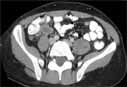 |
CT slice through the mid-abdomen showing multiple normal-appearing organs, which are labeled.
|
 |
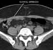
CT scan of a normal appendix in the right lower abdomen. The appendix normally connects with the right colon and contains air (this appears black on the scan). Air in the appendix excludes appendicitis since this means that the appendix is not obstructed or inflamed.
|
 |
Reformatted image of the mid-abdomen creating a "slab" showing the liver and intestines. The abdominal aorta and branches going to the liver, spleen and stomach are visible. This view is looking up toward the head of the patient.
|
 |
Coronal Reconstruction of the Abdomen and Pelvis.
|
 |
CT angiogram. Frontal or coronal view of chest-3D slab image showing pulmonary vessels.
|
 |
CT of the lungs, window level set to demonstrate the vessels and air ways - not intended to demonstrate the heart, spine muscles etc. This is used to look for things like pneumonia or lung cancer.
|
 |
Contrast-enhanced CT of the neck in a patient with lymphoma. Single arrowhead shows abnormal enlarged lymph node in the upper neck on the patient's right side near the angle of the jaw. Double arrowheads show numerous smaller abnormal lymph nodes on the patient's right side.
|
 |
Comminuted Fracture of the left hip on 3D CT Reconstruction.
|
 |
Comminuted Fracture of the left hip on 3D CT Reconstruction 2.
|
 |
Comminuted Fracture of the left hip on 3D CT Reconstruction 3.
|
 |
CT - Normal cross section of the neck just above the vocal cords. This is also known as a transverse or axial image.
|
 |
CT - Normal coronal section of the neck, computer-generated (or "reconstructed") from a series of cross-sections.
|
 |
CT - Normal sagittal section of the neck in the midline, computer-generated (or "reconstructed") from a series of cross-sections.
|
 |
Patient undergoing computed tomography (CT) scan
|
 |
Computed Tomography (CT or CAT scan) equipment
|
 |
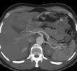
CT scan showing the liver.
|
 |
CT scan of the neck with intravenous contrast. Cross section (also called an axial or transverse section) shows a nodule of the right lobe of the thyroid gland (arrowhead) surrounded by normal thyroid gland, which appears lighter gray in this image.
|
 |
Coronal section, generated from a series of cross sections, shows a nodule of the right lobe of the thyroid gland (arrowhead) surrounded by normal thyroid gland, which appears lighter gray in this image.
|
 |
Computed Tomography (CT) equipment
|
 |


