Images are shown for illustrative purposes. Do not attempt to draw conclusions or make diagnoses by comparing these images to other medical images, particularly your own. Only qualified physicians should interpret images; the radiologist is the physician expert trained in medical imaging.
|
Image Gallery
Lower GI Tract X-ray
|
Click image to view larger |
This image shows the right side of the large intestine. Air (dark) distends the bowel and barium (white) coats the inner lining.
|
 |
X-ray image from a small bowel series showing a right inguinal hernia.
|
 |
Normal air contrast barium enema
|
 |
|
 |
|
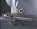 |
X-ray equipment is mounted on a C-shaped gantry with the x-ray tube itself beneath the table on which the patient lies. Above the patient is an image intensifier that receives the x-ray signals, amplifies them, and sends them to a TV monitor.
|
 |
|
|


