|
Click image to view larger |
Plain film radiographic (x-ray) image (anterior-posterior view) of the pelvis in a child, demonstrating sacral agenesis (rare failure of sacral bone development).
|
 |
|
 |
X-ray showing frontal view of both hands.
|
 |
|
 |
Bone Scan - Normal. Two pictures (one from the front and one from the back) of a normal bone scan from a 12-year-old boy.
|
 |
Plain film radiographic (x-ray) image (scaphoid view) of the left wrist, demonstrating fracture of the scaphoid bone.
|
 |
Plain film radiographic (x-ray) image
(posterior-anterior view) of the left hand, demonstrating severe bony destruction and soft tissue swelling of the hand in a patient with longstanding gout.
|
 |
|
 |
Plain film radiographic (x-ray) image (anterior-posterior view) of a normal right foot.
|
 |
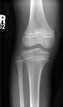
A normal knee in a 12 year old (anterior-posterior view).
|
 |
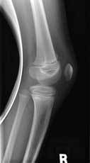
A normal knee in a 12 year old (lateral view).
|
 |
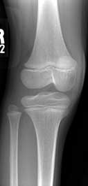
A normal knee in a 12 year old (oblique view).
|
 |
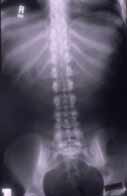
X-ray showing lumbar spine (front view)
|
 |
X-ray showing lumbar spine (side view)
|
 |
Plain film radiographic (x-ray) posterior-anterior image of the left hand, demonstrating a duplicated left thumb.
|
 |
|
 |
Plain film radiographic (x-ray) image (anterior-Posterior view) of a normal cervical spine.
|
 |
Plain film radiographic (x-ray) image (lateral view) of a normal cervical spine.
|
 |
Plain film radiographic (x-ray) image (lateral view) of a normal lumbar spine.
|
 |
This plain film radiographic (x-ray) image (coned lateral view) of the lumbar spine shows the lumbo-sacral junction to better advantage.
|
 |
Radiological technologist preparing to take an arm x-ray on a patient.
|
 |


